Focused Cardiac Ultrasound
-
Welcome
Introduction to FoCUS2 Topics -
Section 1Video: Cardiac Anatomy
-
Cardiac Anatomy3 Topics
-
Section 2Key Video: Obtaining the 5 Views
-
Acquisition & the 4Ps4 Topics|1 Quiz
-
Acquiring The 5 Views5 Topics|1 Quiz
-
Clinical Case Scenario
-
Section 3Interpretation11 Topics|2 Quizzes
-
Interpretation: The RUSH Protocol1 Quiz
-
Section 4Medical Management
-
Foundations of Echocardiography
-
ReferencesReferences & Further Reading
Quizzes
Anatomy Overview
Anatomy
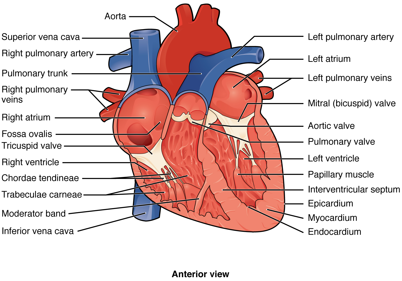
You may take a look at this excellent 3D animated model anatomy overview of the heart. Rotate the image and get an anatomical picture of the heart in your mind. Note the relationship of the 4 heart valves. The pulmonary valve is the most superior valve in the chest. Turn the heart around to visualize the right atrium, superior and inferior vena cava. The left atrium and pulmonary veins draining into it can be seen as well. Play around with the model and get a good feel for the anatomy of the heart and relationship of important structures.


References & Further Reading
An Anatomical Review of the Right Ventricle. Wang et al. Translational Research in Anatomy. 2019
Anatomy of the Mitral Valve. SY Ho. BMJ Heart.
Mitral Regurgitation: Anatomy, Physiology, and Pathophysiology—Lessons Learned From Surgery and Cardiac Imaging. Frontiers in Cardiovascular Medicine. 2020.
Imaging Assessment of the Right Atrium: Anatomy and Function. Lang et al. European Heart Journal. 2022.
An Anatomical Review of the Left Atrium. Whiteman et al. Translational Research in Anatomy. 2019.
Anatomy of the Cardiac Chambers. Review of the Left Ventricle. Whiteman et al. Translational Research in Anatomy. 2021.

