Lung Ultrasound 1
This station covers Lung Ultrasound Basics. We will look at the types of probes, scanning protocols and normal lung ultrasound sonoanatomy.
A. Probe Selection

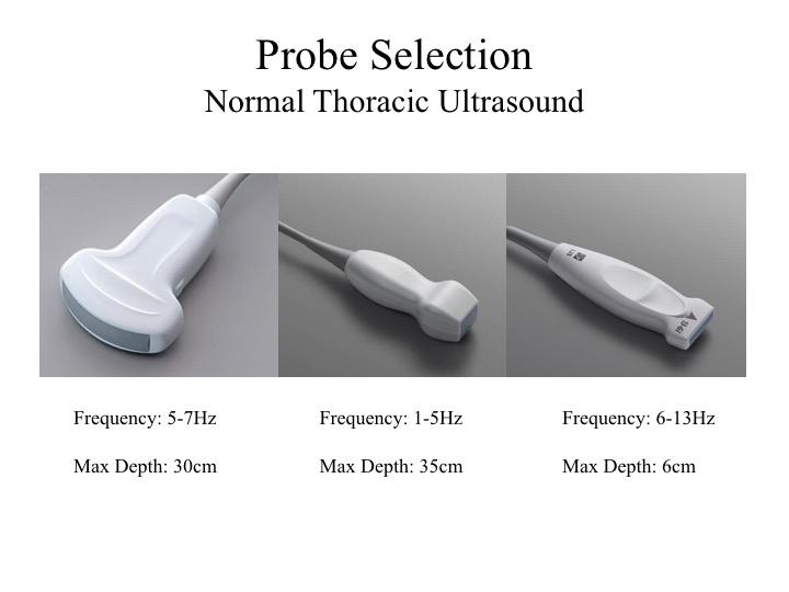
- Linear Probe for visualising the pleural line. High frequency allows for better resolution but shallower penetration (depth).
- Phased Array (Cardiac) or Curvilinear Probe for better penetration and imaging of lung parenchyma. Useful for scanning the lung laterally and posteriorly to look for effusions and consolidation.
- USe Radiology setting for display – marker on the left of the display.
B. Scanning Protocols

1. Eight Zone Protocol
Each hemithorax is divided into 4 areas, to give a total of 8 zones for bothe sides of the chest.
In the supine patient, scan anteriorly when suspecting a pneumothorax and posterolaterally when looking for effusions.
Landmarks:
- PSL: Parasternal Line
- AAL: Anterior Axillary Line
- PAL: Posterior Axillary Line

2. Six Zone Protocol
BLUE Protocol (Bedside Lung Ultrasound in Emergencies):
- Upper BLUE Point
- Lower BLUE Point
- PLAPS Point – Posterior Lateral Alveolar Pleural Point
C. Normal Lung
1. Linear probe
- Linear High Frequency Probe
- Useful for evaluating the pleura and presence or absence of lung sliding.
- Pleura is seen as the bright echogenic line, below the intercostal muscles, between the 2 ribs.
- Presence of lung sliding is normal.
- Seen as shimering / ants crawling
- Lung sliding is present when the parietal and visceral pleura are in apposition, and the lung is ventilated normally.
- Z-lines or comet tails are seen in normal lung. These are short linear artifacts that extend from the pleura for a short distance. They differ from B-lines.
2. Curvilinear Probe
- Curvilinear Low Frequency Probe
- Increased depth acompared to linear probe, but lower resolution.
- Pleural line is seen as an echogenic bright line closest to transducer head.
- A-lines are seen. These are reverberation artifacts caused by reflection of ultrasound waves between the probe head and pleural line.
- A-Lines are present in normal well aerated lungs.
- A-Lines occur at repeatedly at the same distance, which is equal to the distance from the probe head to lung pleura.
3. M-Mode

Seashore Sign: Seen in normal lung sliding.

Barcode Sign: Seen in absence of pleural sliding. Also called the stratosphere sign.
D. Clinical Scenario
A 74 year old man is feeling breathless in PACU following laparoscopic cholecystectomy. His SpO2 has fallen to 93% on air. A lung ultrasound scan is performed. What are the findings?
Left Lung
Left Anterior
Left Posterior Lateral
Right Lung
Right Anterior
Right Posterior Lateral
In the anterior zones, there is normal lung sliding bilaterally. The pleural line is normal. The posterior lung zones show normal well aerated lungs, with the spine disappearing on inspiration as the diaphragm is pushed into the abdomen. The lung scan is normal.

